16 December 2020 : Animal Research
Paclitaxel Ameliorates Palmitate-Induced Injury in Mouse Podocytes
Seung Seob Son1ABCDEF, Jeong Suk Kang12CDEF, Eun Young Lee123ACDEFG*DOI: 10.12659/MSMBR.928265
Med Sci Monit Basic Res 2020; 26:e928265


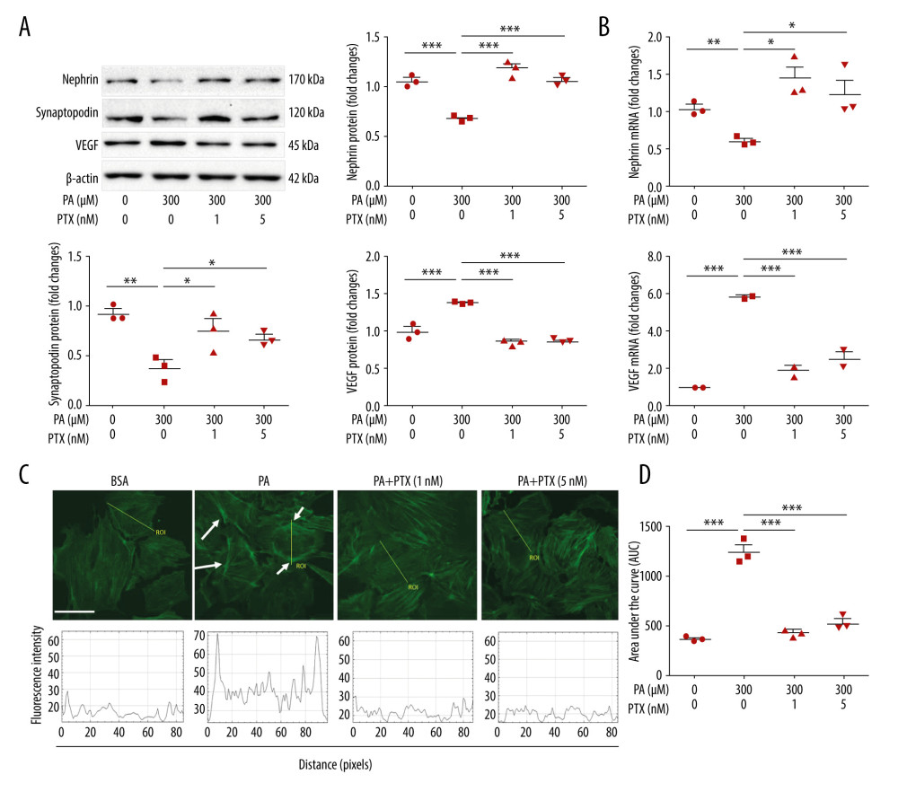
Figure 2 Effect of paclitaxel treatment on palmitate-induced podocyte damage. Mouse podocyte cells were treated with 300 μM palmitate with or without paclitaxel for 24 h. (A) Western blots and densitometry for nephrin, synaptopodin, and VEGF. (B) Real-time PCR for nephrin and VEGF was normalized by β-actin mRNA level in the same sample. (C) FITC-phalloidin staining showing F-actin. Arrows indicate reorganization of F-actin. Line ROC analyses of the fluorescence intensity for each picture are shown at the bottom. Quantification was performed as in [10]. (D) Area under the curve analysis of the results shown in (C). Error bars represent the mean±SEM (n=3). Magnification 40×; bar=60 μm. BSA – bovine serum albumin; PA – palmitate; PTX – paclitaxel: ROC – receiver operating characteristic. * P<0.05, ** P<0.01, *** P<0.001 compared with palmitate.